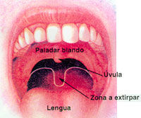
Model of the palate
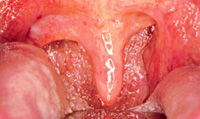
Strong hypertrophy of the uvula
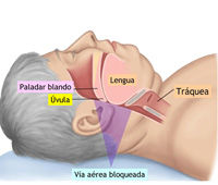
Snoring in adults
Snoring due to a hypertrophic uvula and palate
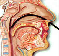
Fibroendoscopy
Snoring in adults
TREATMENT FOR SNORING IN ADULTS WITH/WITHOUT SLEEP APNEA
Snoring can often create problems between couples. If snoring is accompanied by sleep apnoea (a sleep disorder consisting of temporary pauses in breathing) it is not only bothersome but can also be a serious health problem.
This is because sleep apnoea can have many symptoms. When the breathing is suspended, oxygen is prevented from entering the body, which can lead to “obstructive sleep apnoea syndrome” or OSAS.
What are the symptoms?
Snoring mainly affects generally overweight men. In women, the onset of snoring often happens after menopause.
When the sleep apnoea occurs frequently, the patient will experience daytime sleepiness, tiredness and irritability, morning headaches, reduced libido (sexual desire) and high blood pressure.
How is it detected?
Snoring inevitably gets detected when the patient shares a bed with their partner, even more so if the snoring is accompanied by sleep apnoea (pauses in breathing during sleep).
To ensure accurate diagnosis, basic screening is performed, which involves a fibroendoscopy of the nose and throat in order to determine the location of the obstruction causing the problem.
In some cases, a sleep study or polysomnography might also need to be performed, which analyses the intensity of the snoring, the frequency and duration of the pauses in breathing and the quality of the sleep.
How is it treated?
Both sleep apnoea and snoring in adults can be treated in various ways, depending on the criteria established by the ENT (ear, nose and throat) specialist after examining the patient.
- If the patient is overweight, weight loss is essential;
- In many cases, the obstruction is caused by the restriction of the upper airway, i.e. the space between the palate, the uvula, the tonsils and the tongue, due the enlargement of one or more of these structures. In some case, the laser removal of the uvula and part of the soft palate is advisable;
- In some cases, the patient may need to sleep wearing a CPSP nasal mask that prevents the obstruction of the airway by administering a current of air;
- The most common cause of nasal obstruction or blockage is the enlargement (hypertrophy) of the nasal turbinates, as their excessive size makes it difficult for the patient to breathe through the nose.
What are nasal turbinates?
Nasal turbinates are long tube-shaped structures that are found in the lower part of the nose.
They are made up of skin and have bone on the inside.
They have the following functions:
- cleansing
- creating moisture
- heating the air we breathe so that by the time it reaches the throat and lungs it is treated.
The vast majority people who have difficulty breathing through their nose have enlarged nasal turbinates.
- In many cases, the cause is unknown;
- In other cases, they are patients with allergic rhinitis (hay fever). Often, the nasal turbinates grow longer and become increasingly inflamed with age;
- Lastly, the nasal turbinate can become enlarged due to excessive use of nasal sprays or drops, sometimes to such an extreme that the patient is unable to sleep without applying them.
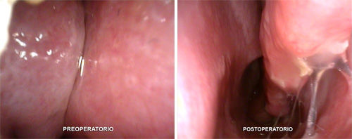
Clinical case
By using an endoscopy technique, we can access the inside of the nose and get a clear view of its interior.
The image to the left shows the lower nasal turbinates that were causing sleep apnoea and snoring (note that the airway is blocked) before surgery. The image to the right is after surgery, where you can see that the airway is no longer blocked and that the space for breathing is much larger.
In the image to the left, we can see that the turbinate is very enlarged and that it hardly leaves space for breathing, resulting in the nasal obstruction, which in turn causes sleep apnoea and snoring.
- Many techniques for reducing nasal turbinates exist, such as radiofrequency However, in a case like this, where the turbinates are very large, the appropriate technique is laser-assisted reduction.
- We can see that the large space that exists after surgery (right-hand image) is a kind of “highway” for the breath. The reduction will last for the rest of the patient’s life. The nasal turbinates will never grow back and the sleep apnoea and snoring can be considered treated.


 by Develona